Fundus Autofluorescence
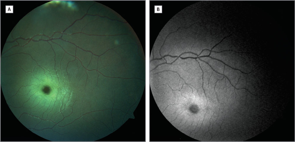
Fundus Autofluorescence (FAF) is a non-invasive retinal imaging modality used to provide a density map of a substance called lipofuscin in the retinal pigment epithelium.
Some natural structures in the retina and optic nerve emit light after they have absorbed light. Autofluorescence in the retinal pigment epithelium (RPE) can be attributed to lipofuscin. Lipofuscin accumulates in the RPE due to age and with certain metabolic defects. This natural emission of light (autofluorescence) can be observed by a special camera or SLO (scanning laser ophthalmoscope). Fundus autofluorescence imaging is a noninvasive technique which helps to discover and delineate changes in retinal structures. Imaging of fundus autofluorescence is used to detect and monitor the progression of various disease states including macular degeneration, diabetic retinopathy, congenital retinal dystrophies, and certain retinal neoplasms.
Fundus Autofluorescence (FAF) imaging in recent years has been shown to be useful in various retinal diseases with regard to diagnostics, documentation of changes, identification of disease progression, and monitoring of novel therapies. This test can help the doctor achieve a better understanding of the health of the fundus in order to obtain earlier diagnoses and better predict the progression of certain retinal diseases.
Call us today at
(561) 322-3588 or Email us at scheduling@specialtyretina.com
Improving Vision • Improving Life